Getting Ready For Cataract Surgery
Before undergoing cataract surgery, it is necessary to measure various parts of the eye, including its length and front curvature. This is necessary for two reasons:
- In order to determine the type and power of lens implant to be placed in the eye, the goal being to choose the lens that will provide the best possible focus to the eye. Most people would like to have the best distance vision without glasses, however some people will choose to see better at near without glasses. Others choose a lens that provides distance and near vision without glasses. There are many options and these need to be customized for each individual. The goal for each patient is determined at the preoperative consultation.
- The measurements obtained allow the surgeon to plan the most successful procedure based on the anatomy of the eye. For example, a very short eye will require a different approach than a very long eye.
- Preoperative eye measurements will determine whether there is any astigmatism, for those patients who wish to improve the focus of their eye by having it surgically corrected at the time of the cataract surgery. Astigmatism means that the eye is football-shaped. The prescription in the glasses does not give us this information. Astigmatism can be improved by implanting a Toric lens.
There are two types of technologies for measuring the eyes: the ultrasound-based method, and the light-based technology (Zeiss IOL Master). The IOL Master uses an infrared laser (wavelength 780 nm) linked to a computer to scan and analyze the dimensions of the eye.
The advantages of the IOL Master are:
- It is more precise than ultrasound. Ultrasound involves the use of sound waves, while laser involves the use of light waves. The more precise the measurements are before the surgery, the better the choice of lens implant power will be, and as a result the better the focus of the eye after surgery.
- It is automatic, which means it removes the human element of holding a probe and aligning it with the eye as is done with the ultrasound method. This eliminates a potential source of error.
- It is faster than ultrasound. This is very helpful for patients with physical conditions that make it difficult for them to stay still during measurement.
- It gives information about the depth of the front part of the eye. This information is very valuable in creating a surgical plan.
- It provides a measurement of astigmatism. This allows the surgeon to plan the best surgical treatment for the astigmatism, if desired. It helps to select the optimal lens style and power by using computer analysis.
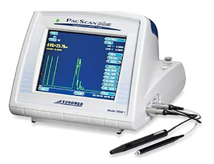
Ophthalmic ultrasound device for measuring the eye.
iTrace For Measuring the Path of Light in the Eye
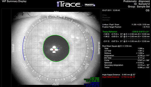
The iTrace measures the quality of vision and visual function using a fundamental thin beam principle of optical ray tracing, a first in eye care diagnostics.
The iTrace sequentially projects 256 near-infrared laser beams into the eye to measure forward aberrations, processing data point-by-point.
This 5-in-1 system provides auto-refraction, corneal topography, ray tracing aberrometry, pupillometry and auto-keratometry.
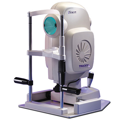
iTrace Wavefront Aberrometer
Optical Alignment - The iTrace helps to determine if a multifocal lens implant would be appropriate for the visual system of the eye.
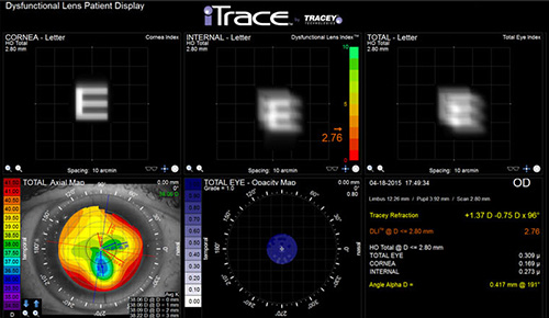
Separate Cornea and Lens - By determining how light travels in the eye, the iTrace provides important information to allow the best choice of lens implant for each eye.
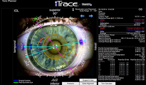
Toric Precision - With iTrace, there is improved precision when using Toric Lens Implants compared to traditional measurement methods.
OCT For Analysing the Various Structures of the Eye
Optical Coherence Tomography (OCT) is performed as required to analyse the various structures of the eye. This test is like an ultrasound except that instead of using sound waves, it uses an infrared laser beam.
Without touching the eye, it provides images of the back of the eye (retina) as well as the front of the eye (cornea). This is the same technology used by the IOLMaster for measuring the dimensions of the eye. It provides very fine detail, down to the range of microns.
In addition to the examination by the ophthalmologist, this scan provides important information about the health of the eye as well as providing information that is important in planning cataract surgery.
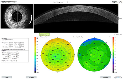
An example of mapping of the front of the eye.
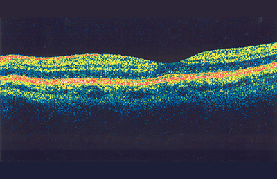
Normal retina.